


肛門管上皮内腫瘍(anal intraepithelial neoplasia:AIN)は、肛門管扁平上皮から移行上皮にかけて発生する比較的稀な腫瘍の総称で、肛門扁平上皮癌の前駆病変とされています。
主としてヒトパピローマウイルス(HPV)が発生に関与するとされ、高リスク型のHPV感染により異型上皮が発生した後、上皮内癌,進行癌へと進展していきますが 、肛門管扁平上皮癌側からみると9割がHPV陽性であるとの報告もあります1.2)。
内視鏡像は隆起型が多く、他に乳頭状、鶏冠状、扁平隆起型などがあり、ルゴール染色では不染帯を呈します。
Narrow band imaging(NBI拡大観察)では、食道異形上皮に類似したドット状・ループ状血管、分布不均一なIPCL(intraepitherial papillary capillary loop)様血管などを示し、範囲や深度の決定に有用である可能性が示唆されています3.4.5)
病理組織診断では、p16やKi67の免疫染色が有用で、p16は高リスク型のHPVに関連した病変で高率に発現し、免疫染色ではhigh grade AINの70〜100%でびまん性に強陽性を示すと報告されていて6)、
WHO分類5版においても、p16染色でブロックパターンに染色されるAIN2/AIN3は、浸潤癌に移行する可能性が高いhigh-grade squamous intraepithelial neoplasia(HSIL)に分類されており、治療対象となります7)。
治療法については、アブレーション、EMR、ESD、トリクロロ酢酸外用、イミキモド外用、フルオロウラシル外用などが有効との報告があり、治療介入により発がん率の抑制効果が示唆されています8)。
最近では、転移のリスクが非常に低いと考えられている上皮内癌やAINに対する内視鏡的治療アプローチが可能になっています9-12)。
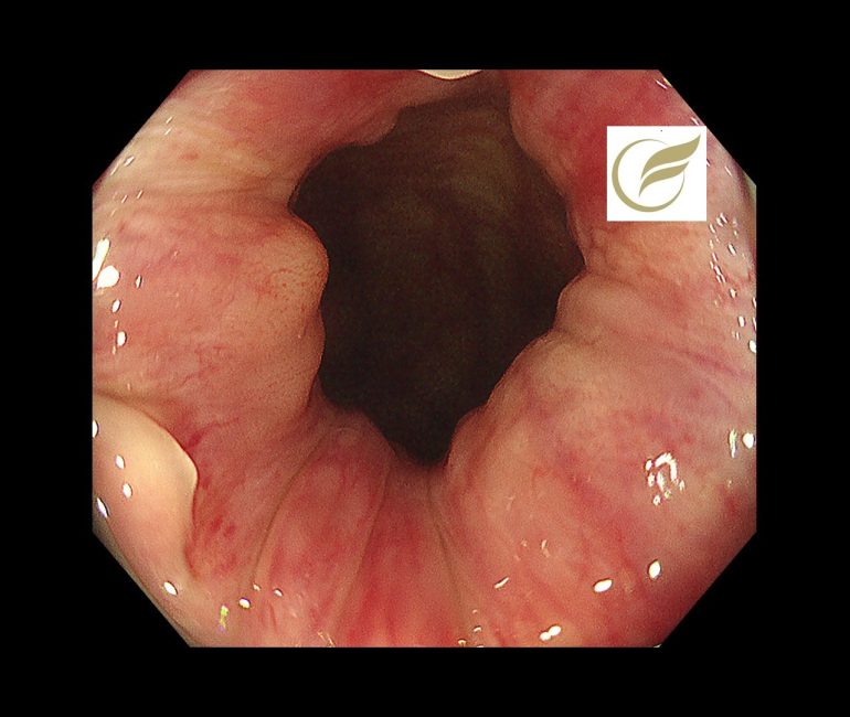
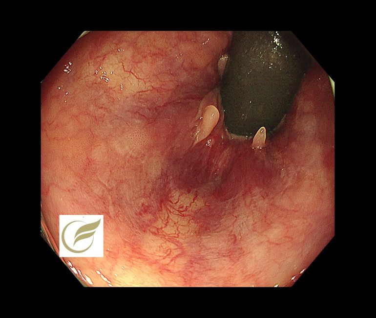
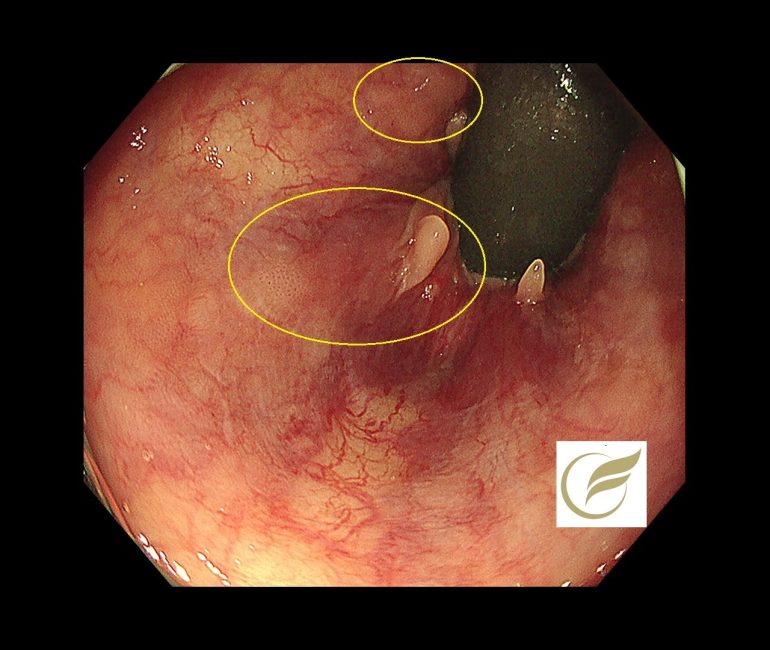
以下は当院で発見されたAINで、小さな扁平隆起の病変と、広い平坦病変の2病変を合併した例です。生検診断で2病変ともAINの診断となりました。

参考文献
- 1) Darragh TM, Colgan TJ, J Thomas Cox JT et al:The Lower Anogenital Squamous Terminology standarzation Project for HPV-Associated Lesions. Arch Pathol Lab
Med, 136:1266-1297,2012]
2) Muñoz N, Bosch FX, de Sanjosé N, et al:Epidermiologic classification of human papillomavirus types associated with cervical cancer. N Eng J Med, 348:518-27,2003
3) Kuwano H, Nishimura Y, Oyama T, et al. Guidelines for diagnosis and treatment of carcinoma of the esophagus April 2012 edited by the Japan Esophageal Society. Esophagus. 2015 Jan; 12(1): 1-30. [DOI] [PMC free article] [PubMed] [Google Scholar]
- 4) Ishihara R, Mizusawa J, Kushima R, et al. Assessment of the diagnostic performance of endoscopic ultrasonography after conventional endoscopy for the evaluation of esophageal squamous cell carcinoma invasion depth. JAMA Netw Open. 2021 Sep; 4(9): e2125317. [DOI] [PMC free article] [PubMed] [Google Scholar]
- 5) J Anus Rectum Colon. 2022 Apr 27;6(2):92-99. doi: 10.23922/jarc.2021-077. eCollection 2022. Anal Intraepithelial Neoplasia: Precursor of Anal Squamous Cell Carcinoma https://pmc.ncbi.nlm.nih.gov/articles/PMC9045852/
6) 高橋雅恵,堀口慎一郎,山澤 翔,他:肛門部尖形コンジローマおよび高異型度肛門上皮内腫瘍の併存例.診断病理,32:136-140,2015
7) Darragh TM, Colgan TJ, Cox JT, et al. The Lower Anogenital Squamous Terminology Standardization Project for HPV-Associated Lesions: background and consensus recommendations from the College of American Pathologists and the American Society for Colposcopy and Cervical Pathology. Arch Pathol Lab Med. 2012 Oct; 136(10): 1266-97. [DOI] [PubMed] [Google Scholar]
8) N Engl J Med . 2022 Jun 16;386(24):2273-2282. doi: 10.1056/NEJMoa2201048. Treatment of Anal High-Grade Squamous Intraepithelial Lesions to Prevent Anal Cancer https://pubmed.ncbi.nlm.nih.gov/35704479/
9)Chou YP, Saito Y, Matsuda T, et al. Novel diagnostic methods for early-stage squamous cell carcinoma of the anal canal successfully resected by endoscopic submucosal dissection. Endoscopy. 2009 Oct; 41(Suppl 2): E283-5. [DOI] [PubMed] [Google Scholar]
- 10) Oono Y, Fu K, Nakamura H, et al. Narrowband imaging colonoscopy with a transparent hood for diagnosis of a squamous cell carcinoma in situ in the anal canal. Endoscopy. 2010 Dec; 42(Suppl 2): E183-4. [DOI] [PubMed] [Google Scholar]
- 11) Kasuga K, Saito Y, Wu SYS, et al. Impact of endoscopic submucosal dissection of an anal squamous intraepithelial lesion with indistinct border. Endoscopy. 2020 Feb; 52(2): E75-7. [DOI] [PubMed] [Google Scholar]
12)Clinical application of endoscopic submucosal dissection for superficially invasive squamous cell carcinoma/high-grade squamous intraepithelial lesion involving the canal anal. Tech Coloproctol. 2024 Jul 31;28(1):90. doi: 10.1007/s10151-024-02966-8.PMID: 39085740
文責 院長 岡田和久